Leg Bone Diagram - Anatomy Chart Of Human Bones For Medicine Design Stock Vector Illustration Of Orthopedics Label 116682162
Leg Bone Diagram - Anatomy Chart Of Human Bones For Medicine Design Stock Vector Illustration Of Orthopedics Label 116682162. Now let's look at the tibia bone, which is the larger of the two leg bones, located medially. The bones of the leg are the femur, tibia, fibula and patella.the foot bones shown in this diagram are the talus, navicular, cuneiform, cuboid, metatarsals and calcaneus. Blank leg bones diagram : The major bones of the leg are the femur (thigh bone), tibia (shin bone), and adjacent fibula, and these are all long bones.the patella (kneecap) is the sesamoid bone in front of the knee.most of the leg skeleton has bony prominences and margins that can be palpated and some serve as anatomical landmarks that define the extent of the leg. He leg's main function in the human is for locomotion and support of the rest of the body.
ads/bitcoin1.txt
Foot and leg bones diagram best secret wiring diagram. This area is commonly referred to as the calf. Depending on the origin of the discomfort, upper leg pain symptoms can be a chronic nuisance or acute and debilitating. Pelvic bone labeled 12 photos of the pelvic bone labeled pelvic bone labeled, pelvic bone labeling quiz. At the same time, the bones and joints of the leg and foot must be strong enough to support the body's weight while remaining.
X Ray Lower Third Leg Bones With Ankle Joint Anteroposterior And Download Scientific Diagram from www.researchgate.net Some types of leg pain can be traced to problems in your lower spine. Printable human skeleton diagram u2013 labeled unlabeled and. Depending on the origin of the discomfort, upper leg pain symptoms can be a chronic nuisance or acute and debilitating. These landmarks are the anterior superior iliac spine. Leg bone stock illustrations joint knee blue icon on white background illustration of joint knee blue icon on white background leg bone stock illustrations The upper leg, in particular, is comprised of bones and muscles that are susceptible to injury, particularly when excess strain is placed upon them. They support bones, in this case, the vertebrae. The rounded, proximal end is the head of the femur, which articulates with the acetabulum of the hip bone to form the hip joint.
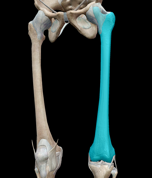
Now let's look at the tibia bone, which is the larger of the two leg bones, located medially.
ads/bitcoin2.txt
Pelvic bone labeled 12 photos of the pelvic bone labeled pelvic bone labeled, pelvic bone labeling quiz. The tibia, commonly known as the 'shin bone', is the largest and most medial of the two.you can palpate its anterior border when you run your finger down the anterior aspect of your leg. Related posts of leg bones anatomy diagram structure of anatomy leg and foot. A long bone has a shaft and 2 ends. The hip itself is a ball and socket joint, much like the shoulder.the structures necessary to create this joint are the socket, the joint capsule, muscle, ligaments, and the neck. The bones of the hip include the femur, the ilium, the ischium, and the pubis. Isolated femur, patella, fibula, tibia and foot bones with shown injury location. The lower leg is comprised of two bones, the tibia and the smaller fibula. Michael sparacino answered 37 years experience family medicine The lower leg extends from the knee to the ankle. Is my extra bone causing this? I've started getting pins and needles up and down left leg, painful. Every skeletal muscle has three main parts:
The bones of the leg are the femur, tibia, fibula and patella.the foot bones shown in this diagram are the talus, navicular, cuneiform, cuboid, metatarsals and calcaneus. Stress fractures of the lower leg are most commonly seen in sports that place repetitive stress on the leg, such as running and jumping sports (e.g., gymnastics, basketball, and tennis). These muscles work together to produce movements such as standing, walking, running, and jumping. The major bones of the leg are the femur (thigh bone), tibia (shin bone), and adjacent fibula, and these are all long bones.the patella (kneecap) is the sesamoid bone in front of the knee.most of the leg skeleton has bony prominences and margins that can be palpated and some serve as anatomical landmarks that define the extent of the leg. Its lower end helps create the knee joint.
3d Skeletal System 5 Cool Facts About The Femur from www.visiblebody.com The rounded, proximal end is the head of the femur, which articulates with the acetabulum of the hip bone to form the hip joint. The tibia, commonly known as the 'shin bone', is the largest and most medial of the two.you can palpate its anterior border when you run your finger down the anterior aspect of your leg. These landmarks are the anterior superior iliac spine. There are in all 7 bones, which fall under tarsal bones category. This area is commonly referred to as the calf. Basically, the muscles surrounding bone become so fatigued from overuse that they eventually transfer the stress onto the bone, leading to a tiny break. It also separates muscles on the anterior and posterior parts of the leg. The lower leg is comprised of two bones, the tibia and the smaller fibula.
A long bone has a shaft and 2 ends.
ads/bitcoin2.txt
The bones of the leg are the femur, tibia, fibula and patella.the foot bones shown in this diagram are the talus, navicular, cuneiform, cuboid, metatarsals and calcaneus. Depending on the origin of the discomfort, upper leg pain symptoms can be a chronic nuisance or acute and debilitating. The knee joint is the largest joint in the body and is primarily a hinge joint, although some sliding and rotation occur. A long bone has a shaft and 2 ends. The tibia, commonly known as the 'shin bone', is the largest and most medial of the two.you can palpate its anterior border when you run your finger down the anterior aspect of your leg. These muscles work together to produce movements such as standing, walking, running, and jumping. By tightening and relaxing, the skeletal muscles create movement. The tibia and fibula are two long bones that run parallel to each other, forming the scaffold of the leg and providing attachment points for many muscles. Foot and leg bones diagram best secret wiring diagram. Stress fractures of the lower leg are most commonly seen in sports that place repetitive stress on the leg, such as running and jumping sports (e.g., gymnastics, basketball, and tennis). *the origin, insertion, and belly.* a muscle's origin is where a tendon attaches it to the *less* movable bone. The tibia and the fibula, at the top of the ankle joint. (note, the radius and ulna bones also have this membrane.) this membrane keeps the tibia and fibula together and provides strength and stability for them.
Is my extra bone causing this? The lower leg extends from the knee to the ankle. Michael sparacino answered 37 years experience family medicine Start studying lab test 2: The tibia, commonly known as the 'shin bone', is the largest and most medial of the two.you can palpate its anterior border when you run your finger down the anterior aspect of your leg.
Muscles Of The Leg And Foot Classic Human Anatomy In Motion The Artist S Guide To The Dynamics Of Figure Drawing from doctorlib.info The bones together make up the hip. He leg's main function in the human is for locomotion and support of the rest of the body. The diagram of bones in the ankle and foot is given below: Learn vocabulary, terms, and more with flashcards, games, and other study tools. A long bone has a shaft and 2 ends. The tibia and fibula are two long bones that run parallel to each other, forming the scaffold of the leg and providing attachment points for many muscles. Leg bone anatomy diagram diagram of human leg human anatomy diagram 10 / 10 ( 1 vote ) in this image, you will find femur, medial epicondyle of the femur, patella, tibial tuberosity, anterior rest of the tibia, a medial surface of the tibia, lateral epicondyle of the femur, head of the fibula, fibula, medial malleolus of the tibia, lateral. Related posts of leg bones anatomy diagram structure of anatomy leg and foot.
The tarsal bones in the foot are located amongst tibia, metatarsal bones, and fibula.
ads/bitcoin2.txt
They support bones, in this case, the vertebrae. By tightening and relaxing, the skeletal muscles create movement. The tibia and fibula are two long bones that run parallel to each other, forming the scaffold of the leg and providing attachment points for many muscles. Now let's look at the tibia bone, which is the larger of the two leg bones, located medially. Depending on the origin of the discomfort, upper leg pain symptoms can be a chronic nuisance or acute and debilitating. Michael sparacino answered 37 years experience family medicine Our goal is that these leg anatomy worksheets pictures gallery can be a direction for you, bring you more references and also make you have a great day. Related posts of diagram of leg bones pelvic bone labeled. Printable human skeleton diagram u2013 labeled unlabeled and. A long bone is a bone that has greater length than width. Blank leg bones diagram : This area is commonly referred to as the calf. The femur, or thigh bone, is the single bone of the thigh region (figure 6.51).
ads/bitcoin3.txt
ads/bitcoin4.txt
ads/bitcoin5.txt
0 Response to "Leg Bone Diagram - Anatomy Chart Of Human Bones For Medicine Design Stock Vector Illustration Of Orthopedics Label 116682162"


0 Response to "Leg Bone Diagram - Anatomy Chart Of Human Bones For Medicine Design Stock Vector Illustration Of Orthopedics Label 116682162"
Post a Comment